Frequently Asked Questions
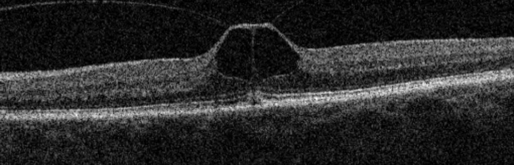
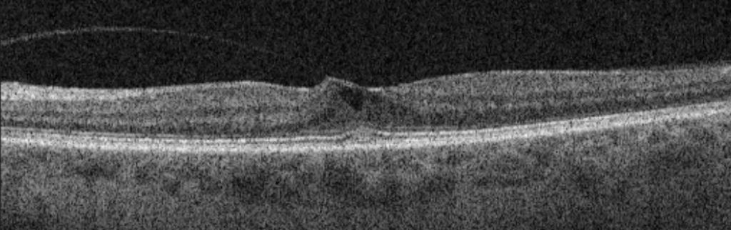
What is vitreo-macular traction (VMT)?
The natural ageing process in the jelly of the eye usually results in a posterior vitreous detachment (PVD). In some cases, vitreous jelly separation from the retina is incomplete and there are parts of the jelly that remain stuck at the centre of the retina. These adhesions can exert a pull on the retina and cause tenting or lifting of some or all of the retinal layers. Sometimes this produces no symptoms and is picked up incidentally on a routine OCT scan of the retina. In other cases, blurring of vision, scotoma (blind spot) or distortion symptoms may result.
What treatment is required for VMT?
VMT often remains relatively stable over time. A proportion can progress to worsening retinal distortion or occasionally a full thickness macular hole. Monitoring with OCT scans is usually advised for a period of time to establish that the retinal appearance is not changing.
If the VMT is not producing any symptoms, no treatment is indicated. We may still monitor things for a few months to see if any changes are occurring. Mild visual problems may also not require treatment, as surgical intervention in some of these cases is not guaranteed to bring about visual improvement.
If the symptoms are significant or if the VMT is seen to be getting worse, surgery may be offered. The operation for VMT is called a vitrectomy, often combined with an inner limiting membrane (ILM) peel.
An injection treatment for VMT is also available, with a medication called Ocriplasmin®. This is designed to try to dissolve the strands of vitreous gel which are stuck to the retina and release the pulling force. If this treatment is appropriate in your case, it will be discussed and offered during your consultation.
What happens during surgery?
A vitrectomy procedure takes around from 30 minutes – 1 hour and is done as a day case procedure. You will have some eye drops put in before surgery in order to dilate the pupils. A local anaesthetic will be administered, usually in the form of an injection of anaesthetic next to the eye. A mild sedative may be administered to allow you to feel more relaxed. We ask you to lie down on a bed and ensure that you are comfortable for the procedure. The area around the eye will be cleaned with an antiseptic solution and a tented cover placed over half of the face to ensure cleanliness and comfort.
A keyhole approach is used to remove the vitreous jelly inside the eye to gain access to the retina. The vitreo-macular traction is released and a full retinal examination is performed. After surgery, it is common to leave a bubble of air inside the eye. This will impair your vision for several days while the air bubble is slowly dispersing.
After surgery, it is necessary to use regular eye drops for several weeks to control inflammation and help prevent infection. We ask you to use a plastic protective shield around the eye at night time for a couple of weeks to prevent accidental injury.
Most operations proceed very smoothly, however sometimes if some other eye conditions or complicating features are present, the operation time and/or the recovery can be prolonged. If this applies to you, the surgeon will discuss it with you before and after your operation and answer any questions you may have.