Frequently Asked Questions
What is an epiretinal membrane?
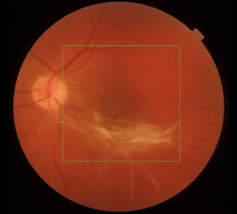
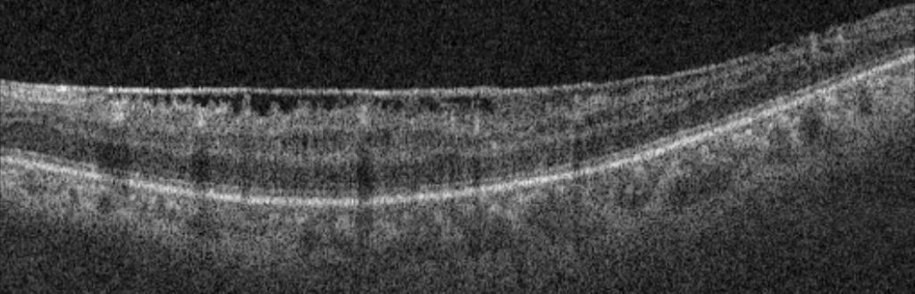
The retina is a thin “wallpaper” inside the eye. It is comprised of layers of nerve tissue and acts like the photographic film in a camera, picking up the light that enters the eye and sending images to the brain. The central part of the retina is called the macula and this is responsible for the sharp vision we use when reading and recognising faces. Sometimes, fibrous scar tissue called an epiretinal membrane (ERM) can grow across the macula. This is a common condition associated with ageing changes in the eye. Epiretinal membranes are also more likely to occur following inflammation in the eye, injuries, or eye surgery. It has been observed in about 10% of people and in most cases does not give rise to any symptoms. It is likely to remain stable over time and usually does not require regular observation.
What problems does an ERM cause?
Most patients will not notice a significant visual problem from an ERM. Occasionally, if the fibrous tissue of the ERM thickens and contracts, it can start to twist and crinkle the retina. In this case, some people can become aware of blurred vision and distortion, where images look bent out of shape.
How are ERMs treated?
In the absence of any symptoms, no treatment is indicated for epiretinal membrane. If you have an epiretinal membrane which is causing visual problems, surgical treatment is available. The operation is called a vitrectomy and epiretinal membrane peel.
A vitrectomy and ERM peel procedure takes anywhere from 30 minutes to 1 hour, depending on complexity. It is usually done as a day case procedure. You will have some eye drops put in before surgery in order to dilate the pupils. A local anaesthetic will be administered, usually in the form of an injection of anaesthetic next to the eye. A mild sedative may be administered to allow you to feel more relaxed. We ask you to lie down on a bed and ensure that you are comfortable for the procedure. The area around the eye will be cleaned with an antiseptic solution and a tented cover placed over half of the face to ensure cleanliness and comfort.
A keyhole approach is used to remove the vitreous jelly inside the eye and gain access to the retina. The epiretinal membrane is carefully removed from the retina and a full retinal examination is performed. After surgery, it is common to leave a bubble of air inside the eye. This will impair your vision for several days while the air bubble is slowly dispersing. As the vision returns, it is normal for a little blurring and distortion to persist. Vision continues to improve slowly over several months following surgery.
After surgery, it is necessary to use regular eye drops for several weeks to control inflammation and help prevent infection. We ask you to use a plastic protective shield around the eye at night time for a couple of weeks to prevent accidental injury.
Most operations proceed very smoothly, however sometimes if some other eye conditions or complicating features are present, the operation time and/or the recovery can be prolonged. If this applies to you, the surgeon will discuss it with you before and after your operation and answer any questions you may have.
What will my vision be like after surgery?
It is important to appreciate that some visual after effects such as mild blurring or a little residual distortion will persist after surgery. These continue to improve for a number of months and occasionally even years post-operatively, as the retina recovers from the effect of having had the ERM.